Pythium is a genus with over 200 species found worldwide, some of which are residents of terrestrial habitats while others are aquatic. In terms of nutrient acquisition, species within the genus may be saprophytes, plant or animal parasites, or mycoparasites. The genus was established by Pringsheim in 1858 (13) and members were considered as true fungi until recently when they were moved to a new Kingdom, Stramenopila. Within that kingdom, the genus Pythium is in the phylum Heterokonta, class Peronosporomycetes, order Pythiales, family Pythiaceae (7). The order Pythiales also includes the genera Phytophthora and Pythiogeton.
Pythium species are eukaryotes (have true nuclei) that have filamentous (thread-like), coenocytic (non-septate threads lacking cross walls) cell growth. The cell wall of many oomycetes is composed of cellulose and β-1, 3 glucan with minimal amounts of chitin. Chitin is a major component of the walls of true fungi. The asexual or vegetative stage of Pythium produces chlamydospores (thick walled resting spores), sporangia (that germinate directly to produce a hypha or indirectly to give rise to vesicle outside the sporangium, within which zoospores are formed), and hyphal swellings (spherical sporangia-like structures that do not give rise to zoospores). During sexual reproduction, an antheridium fertilizes an oogonium to produce a thick walled oospore. Some Pythium species are heterothallic and require opposite mating types to reproduce sexually but most are homothallic and do not require an opposite mating type. Standard keys (17;6) differentiate species within Pythium based on the shapes, sizes, and locations on the mycelium of the sporangium, antheridium, oogonium, and oospore.
Not all species or isolates within some species form zoospores. Zoospores are asexual, bi-flagellated, thin walled spores that move toward host cells where they encyst. The cysts then produce a germ tube and an appressorium that aids in the penetration of the host's cells. The sporangium of some species proliferate. That is, they give rise to additional sporangia either within an existing sporangium or immediately outside a sporangium. Oospores and sporangia are considered the primary survival structures of most Pythium species (11). Oosporogenesis is favored by salinity levels consistent with their normal habitats (eg. water vs. soil) (10) as well as low ratios of carbon/nitrogen typical in infected host tissue.
One way to determine quickly whether the organism you are examining is a Pythium or determine that the pathogen affecting a plant is Pythium is to employ a kit available from Neogen (ALERT-LF®). This kit is easy to use and gives a result in about 20 minutes. A positive test tells you that the organism present is probably in the genus Pythium. There are no kits for identifying the species present. Also note that in rare cases, there can be a cross reaction with Phytophthora. Kits for detecting Phytophthora are available through Agdia, Inc. (Immunostrip®) and Pocket Diagnostic. The Pocket Diagnostic® kit is not licensed for sale in the U.S., but is available in Canada.
When examining hyphae to determine whether you have Pythium, use a light microscope. An inverted light microscope works very well for this when examining agar or liquid in Petri plates. Hyphae of Pythium may have one or more of the following characteristics:
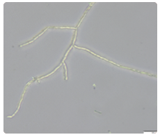
Pythium species have coencytic hyphae (lack cross walls) and hyphae are not dichotomously branched (bottom left). In comparison, dichotomously branched hyphae (bottom right) have hyphal branches that are equal in length, at least at first. Pseudoperonospora, shown in the image (bottom right) (Gerald Holmes, Valent USA Corporation, Bugwood.org), exhibits dichotomous branching. The colored arrows added to the image help to visualize the equal branch lengths.


Hyphae have terminal or intercalary spherical or swollen structures that are part of the hyphal thread, like a bead on a string (right). The images below show examples of sporangia and oogonia. As you can see in the labeled image to the right, sporangia can be differentiated from oogonia by the presence of antheridia.


Also, hyphal swellings exist that are neither spornagia or oogonia, however, it is very difficult to distinguish these swellings from young sporanigia and oogonia when observing these structures in a culture. The labeled image to the right shows the hyphal swellings often produced by P. catenulatum.
Hyphae may have swollen cells, slightly larger than the normal hyphae or much larger than normal hyphae. The images below are examples of inflated sporangium.

Pythium hyphae that are in contact with the edge or bottom of the Petri plate are slightly swollen and curled over forming appressoria (images below).

Cultures may show a pattern on various agars. The cultures below are on water agar.


